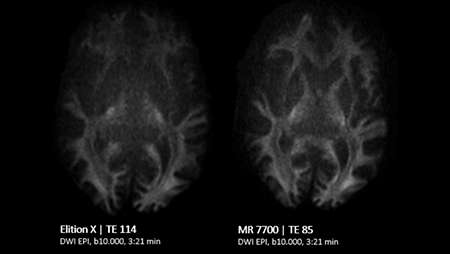
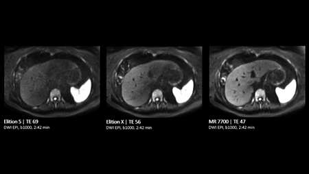
1 Compared to Ingenia Elition X with Vega HP gradients.
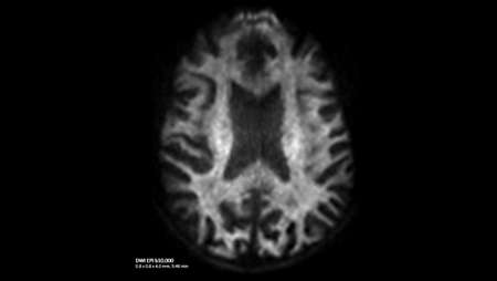
2 Compared to Ingenia Elition X with Vega HP gradients, measured in brain white matter.
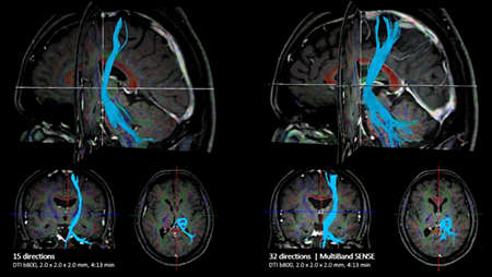
3 Compared to Philips DTI/fMRI scans without MultiBand SENSE.
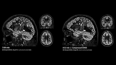
4 Compared to Philips SENSE imaging.
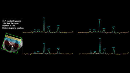
*Caution: Investigational device for imaging with fluorine (19F). Limited by federal (or United States) law to investigational use. Clinical imaging with this nucleus requires usage of a cleared drug. No FDA-cleared drugs are currently available for this nucleus.